The Birthplace of MRI
Stony Brook University is proud to be the home of one of the most groundbreaking medical advancements of the 20th century: Magnetic Resonance Imaging (MRI).
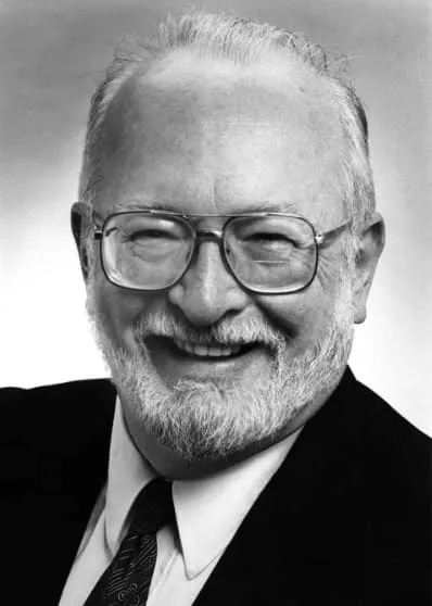
In 1971, Stony Brook researcher Dr. Paul Lauterbur transformed medical imaging by creating the first multi-dimensional image using Nuclear Magnetic Resonance (NMR). His discovery made it possible to see inside the human body without surgery or X-rays, paving the way for the MRI technology we rely on today.
Dr. Lauterbur’s innovative work earned him the 2003 Nobel Prize in Physiology or Medicine, shared with Sir Peter Mansfield.
A Legacy of Innovation
Dr. Lauterbur’s breakthrough came from a simple moment of inspiration: While eating a hamburger, he realized that using a non-uniform magnetic field could create detailed images. This idea became the foundation of modern MRI.
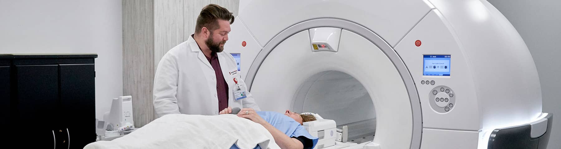
Today, MRI is essential for diagnosing and treating conditions such as cancer and multiple sclerosis. Building on that legacy, Stony Brook physician-scientists also helped pioneer PET imaging and went on to become one of the first hospitals in the United States to offer clinical PET/MRI in 2013.
PET/MRI represents the next step in imaging technology, combining Positron Emission Tomography — which shows how the body functions — with MRI’s detailed anatomical images in a single, simultaneous high-resolution scan. This hybrid approach gives physicians unprecedented clarity to detect, stage and treat disease more quickly and accurately, while using lower doses of radiation.
Continuing the Vision
Today, Stony Brook Imaging and Radiology remains at the forefront of imaging innovation. PET/MRI is used to diagnose and study abdominal and pelvic cancers, neurodegenerative disease, psychological disorders and cardiac conditions, as well as to plan complex surgical procedures.
From Nobel Prize–winning discovery to next-generation hybrid imaging, Stony Brook continues to push the boundaries of biomedical imaging — transforming how we see, understand and treat disease.